
Figure 6.
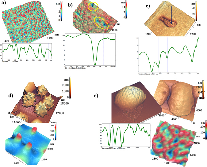
AFM-images of typical nanosurfaces of II order of PRBC membranes and corresponding nanosurface profiles. (a) For discocyte on the 5th day of PRBC storage. (b) Single topological defects (highlighted by yellow circles) on the surface of membranes on the 16th day of PRBC storage. (c) Domains with a grain-like structure.(d) Spheroehinocyte on the 33th day of storage and its nanosurface. (e) Swelled cells e on the 40th day of storage and its nanosurface.