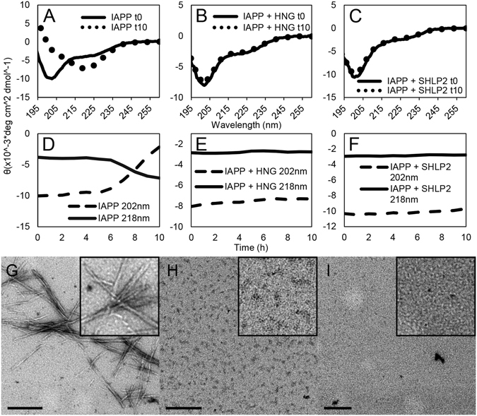
Figure 3.

Time-resolved circular dichroism and transmission electron microscopy confirm MDPs prevent loss of IAPP monomers and prevent fibrilization. (A–C) Circular dichroism spectra of 15 μM IAPP with molar equivalents of MDPs recorded at the beginning and end of each experiment, displayed as a weighted mean residual ellipticity (MRE’). For details regarding the calculation of MRE‘ see Materials and Methods section. (D–F) Time resolved CD of experiments in (a–c). Ellipticities were recorded at 202 nm and 218 nm to follow transitions from random coil (202 nm) to β-sheet (218 nm). Traces represent an average of at least 3 experiments. (G–I) Electron micrographs taken at the end of the experiment for (G) IAPP alone, (H) IAPP treated with HNG, and (I) IAPP treated with SHLP2. Scale bars equal 200 nm.
