
Figure 5.
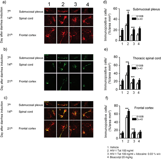
(a–c) Intracolonic administration of HIV-1 Tat caused a marked increase of Cx-43 (green) protein expression in S100B-positive cells (red) in the submucosal plexus, spinal cord and frontal cortex, respectively. (d–f) Quantitative analysis revealed that administration of lidocaine significantly reduced Cx-43 expression on S100B-positive cells of HIV-1 Tat group, whereas bisacodyl did not produce any change in Cx-43 expression compared to vehicle group (S100B (open bars) and Cx-43 (filled bars) expression in the analyzed areas. ***p < 0.001 vs all other groups; °°°p < 0.001 vs HIV-1 Tat group. Scale bars: 20 μm; n = 6 for each group.