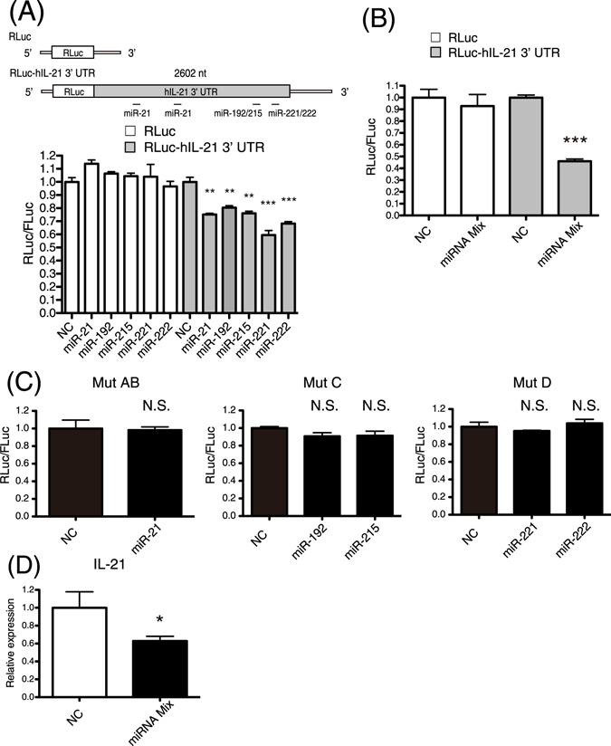
Figure 3.

The expression of human IL-21 was suppressed by miRNAs. (A) 293 T cells were co-transfected with a reporter vector (Renilla or Renilla-hIL-21 3′ UTR), a transfection control vector (pGL3-control), and negative control RNA (NC) or miRNA. Final concentration of NC or miRNAs was 100 nM. Luciferase assays were performed at 24 hours after the transfection. (B) Luciferase assays were performed as described in (A). NC (final 250 nM) or mixture of miR-21, 192, 215, 221, and 222 (final 50 nM each) were co-transfected into 293 T cells. (C) Luciferase assays were performed as described in (A) with reporter vectors with mutant 3′ UTR of human IL-21. (D) NC (final 250 nM) or mixture of miR-21, 192, 215, 221, and 222 (final 50 nM each) were co-transfected into human Th2 cells. Relative expression levels of IL-21 mRNA were examined by quantitative real-time RT-PCR at 24 hours after the transfection. The expression levels were normalized to GAPDH. Data are shown as means + SEM of four independent experiments performed. (A–D) Data shown are a single experiment representative of three independent experiments performed. *P < 0.05, **P < 0.01, ***P < 0.001. N.S., not significant.
