Figure 1.

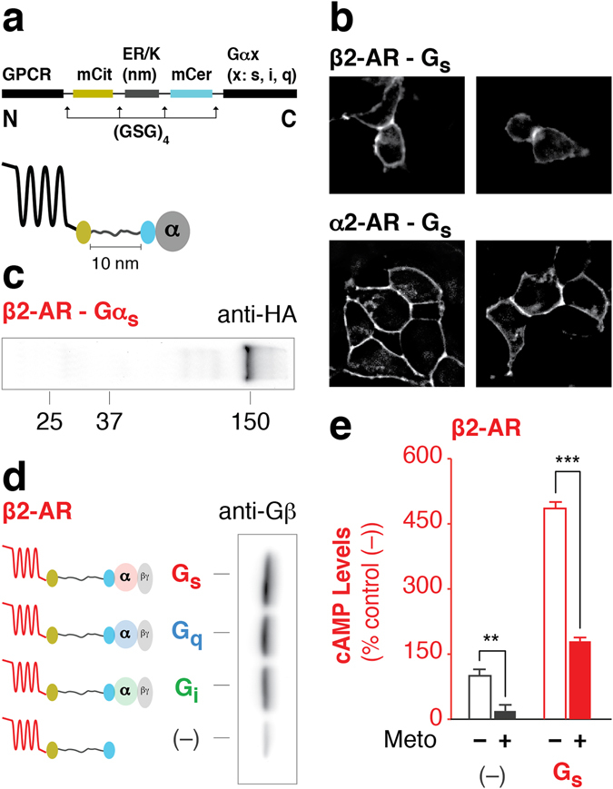
GPCR-G protein fusion sensors are intact and functional. (a) Schematic of the GPCR-G protein sensor design. Protein domains are separated by (GSG)4 linkers to ensure rotational freedom. Control (–) sensors do not contain a Gα subunit. (b) Sensors localize to the plasma membrane in live HEK293T cells as shown in representative images. (c) Western blot of membranes expressing HA-β2-AR-Gαs probed with anti-HA antibody. A distinct 150 kDa band indicates intact sensor expression. (d) Membranes expressing HA-β2-AR-Gαx sensors were subjected to HA-affinity purification and probed with anti-Gβ antibody. Equivalent amount of sensor is loaded per lane as assessed by mCitrine fluorescence. Gβ associates with the Gαs, Gαi, or Gαq subunit. (e) cAMP production in the presence or absence of inverse agonist (100 μM metoprolol) for β2-AR tethered with or without Gs. Data are derived from at least three independent experiments, with at least three replicates per condition. Data are represented as % no-G protein control (–) (mean ± S.E.M Student’s t-test was performed to evaluate significance **p ≤ 0.01 and ***p ≤ 0.001).
