
Figure 2.
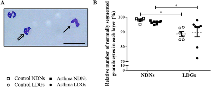
Levels of normally segmented granulocytes in each layer. (A) Representative photography of cytospins (x400, stained Protocol Hema 3) of the peripheral blood mononuclear cell layers (scale bar = 28 µm). Mature granulocytes (empty arrow) have more than 2 nuclear lobes (classically between 3 and 4) connected by filaments, whereas immature granulocytes (full arrow) have a curved nucleus with 2 or fewer nuclear lobes. LDNs were quantitated morphologically by light microscope. (B) Percentages of normally segmented LDNs in peripheral blood mononuclear cells of controls and horses with severe asthma. Each symbol denotes a single animal (mean ± SEM). *p < 0.05 compared with control.