
Figure 6.
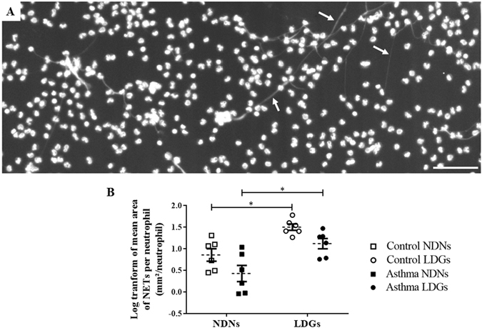
Neutrophil extracellular traps (NETs) production in both layers of cells. (A) Representative photography of using a MRC1024 confocal laser-scanning microscope at ×100 magnification (BioRad, Hercules, CA) equipped with a Nikon Eclipse TE300 camera (Nikon, Tokyo, Japan) of the low-density layer (scale bar = 100 µm). White arrows indicate NETs’ structures. (B) Log transform of the mean area of NETs per neutrophil in each layer (non-stimulated NS). Each symbol denotes a single animal. Mean ± SEM for each studied population is shown. *p ≤ 0.05.