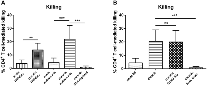
Figure 4.

In vivo cytotoxicity of CD4+ T cells during chronic FV infection. Lymph node cells and spleen cells from naïve mice were loaded with CD4+ T cell FV specific epitope peptides and labeled with CFSE dye. Unloaded cells were stained with cell trace violet. Peptide loaded and unloaded cells were injected intravenously in a 1:1 ratio into naïve and FV-infected mice and tracked by flow cytometry 20 hours after injection. The figure shows the percentage of the target cell killing in the lymph nodes. (A) Cells from naïve mice were loaded either with H2-Ab-restricted F-MulV H19 envelope epitope alone (H19-Env) or with nine additional epitope peptides spanning the coding regions gag, pol, and env (epitope mix), which were described by R.J. Messer et al.33 and injected intravenously into naïve, acutely FV-infected (n = 8), chronically FV-infected (n = 12), chronically FV-infected CD4+ T cell depleted mice (n = 4). (B) Cells from naïve mice were loaded with the peptide epitope mixture. Peptide loaded and unloaded cells were injected intravenously into naïve, acutely FV-infected (n = 8), chronically FV-infected (n = 12), chronically FV-infected GzmB KO mice (n = 8), chronically FV-infected mice, receiving a FasL blocking antibody (n = 4). Mice receiving an isotype control antibody did not show any difference to the non-treated control group. Mean values are indicated by a line. Statistically significant differences between the groups were determined by the unpaired t test: **p < 0.005; ***p < 0.0001, ns, not significant. Data were pooled from two independent experiments.
