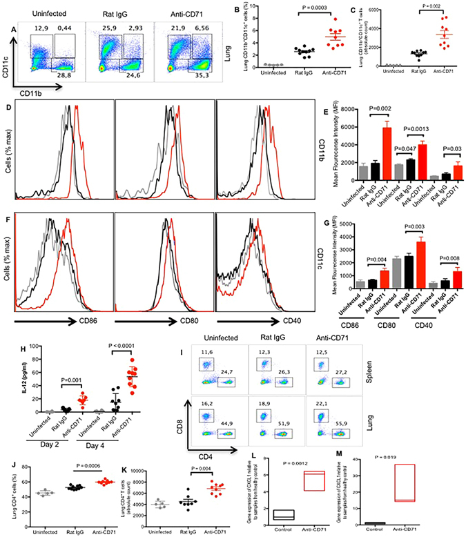
Figure 2.

Deletion of CD71+ cells unleashes recruitment and function of immune cells in the lung in response to B. pertussis low dose infection. (A) Representative dot plots showing percentages of CD11b+, CD11c+ and CD11b+CD11c+ cells in the lungs of newborns day 2 post infection with B. pertussis. (B,C) Percentages and absolute number of CD11b+ and CD11b+CD11c+ cells isolated from the digested lung tissues obtained from the anti-CD71 treated versus isotype control at day 2 post infection. (D–G) Relative CD86, CD80, and CD40 expression by CD11b+ or CD11c+ cells recovered from the lungs at day 2 post infection, treated with anti-CD71 (red histograms gated on CD11b+ or CD11c+cells) or isotype control antibody (black histograms gated on CD11b+or CD11c+ cells) and uninfected mice (gray histograms gated on CD11b+or CD11c+ cells) administered 200 μg of each antibody at day 5. (H) IL-12 levels recovered from the lung homogenate of mice at day 2 and 4 post infection, treated with anti-CD71, isotype control antibody or uninfected mice. (I) Representative plots showing percentages of CD4+ and CD8+ T cells. (J,K) Percentages of CD4+ and CD8+ T cells from the digested lung tissues at day 2 post infection, either treated with anti-CD71 or isotype control compared with each other and uninfected mice. (L,M) CXCL-1 and CXCL-2 genes expression in the lungs of anti-CD71 or isotype control treated mice after B. pertussis infection compared with uninfected mice. Each point represents data from an individual mouse, representative of at least three independent experiments. Bar, mean ± one standard error.
