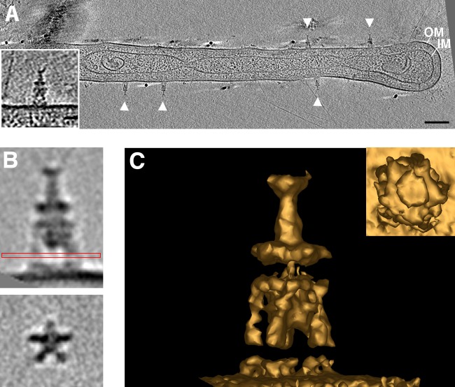
FIG 1.
Novel Prosthecobacter debontii appendages. Multiple external appendages (arrowheads) were observed by ECT on P. debontii prosthecae. (A) A central tomographic slice is shown, with a single appendage enlarged in the inset. Subtomogram averaging revealed the structure in more detail. (B) Side (top) and top (bottom) views show the characteristic disc-like densities and the five legs attaching to the cell surface. The red box shows which view was used to rotate the image 90° for the bottom image. (C) 3D isosurface of the average, seen from the side and top (inset). Bars, 50 nm (A) and 20 nm (inset).

