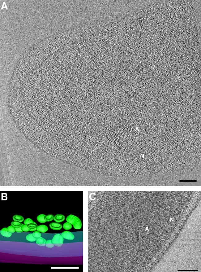
FIG 4.
Novel Vibrio cholerae nanospheres. Clusters of nanospheres were observed in two cryotomograms of V. cholerae cells (central slices shown in panels A and C). “N” indicates nanospheres; “A” indicates associated filament array. (B) Segmentation of the cluster seen in panel A, with outer and inner membranes in magenta and cyan, respectively, and nanospheres in green. A clipping plane cuts through the 3D segmentation revealing the thick walls and hollow centers of the nanospheres. Bars, 100 nm.

