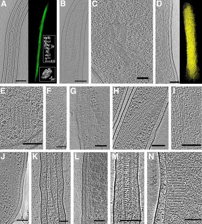
FIG 5.
Filament bundles, arrays, and chains. Hyphomonas neptunium division stalks contained helical bundles (A) that straightened when cells were treated with ethidium bromide (B). The right side of panel A shows a 3D segmentation of the helical bundle, with side and top views of subtomogram averaged insets. Labeled dimensions are in nanometers. (C) Large filament bundles in Helicobacter pylori. (D) A long mesh-like filament array in Vibrio cholerae, with segmentation at right. (E) A more typical V. cholerae filament array. (F to J) Filament arrays in Thiomonas intermedia (F), Hyphomonas neptunium (G), Hylemonella gracilis (H), Halothiobacillus neapolitanus c2 (I), and Mycobacterium smegmatis (J). (K) A chain in Prosthecobacter vanneervenii. (L to M) Filament arrays in Prosthecobacter debontii. (N) A filament array in a starved Campylobacter jejuni cell. Bars, 100 nm (A, B, D to J, L to N) and 50 nm (C and K).

