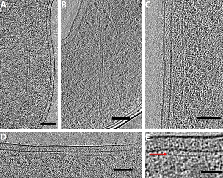
FIG 6.
Single and paired filaments. Tomographic slices showing paired filaments in Campylobacter jejuni (A) and Thiomicrospira crunogena (B) and membrane-aligned filaments in Shewanella putrefaciens (C), Prosthecobacter debontii (D), and Prosthecobacter fluviatilis (E) (the arrow shows a filament just under the inner membrane). Bars, 100 nm (A to D) and 50 nm (E).

