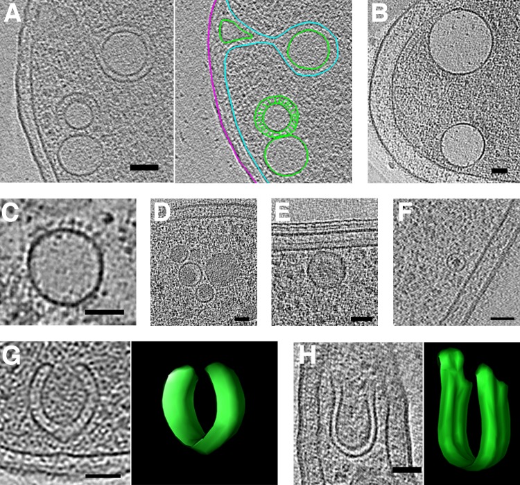
FIG 8.
Round and horseshoe-shaped vesicles. Tomographic slices showing examples of round vesicles in Escherichia coli (A) (segmentation shown at right), Helicobacter pylori (B), Helicobacter hepaticus (C), Myxococcus xanthus (D), Caulobacter crescentus (E), and Myxococcus xanthus overexpressing PilP-sfGFP (F). Examples of horseshoe-shaped vesicles in Ralstonia eutropha (G) and Prosthecobacter fluviatilis (H), with 3D segmentations shown at right. In the segmentation in panel A, outer and inner membranes are in magenta and cyan, respectively, and vesicles are in green. Bars, 50 nm.

