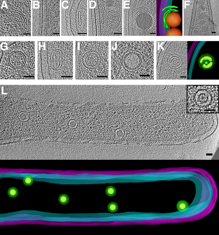
FIG 9.
Flattened and nested vesicles. Examples of flattened vesicles in Thiomonas intermedia (A), Caulobacter crescentus (B to E), and Prosthecobacter debontii (F). Note storage granules in panels E and F, shown in orange in the segmentation panel E (G to L). Examples of nested vesicles in Serpens flexibilis (G), Caulobacter crescentus (H), Borrelia burgdorferi (I), Vibrio cholerae (J), Caulobacter crescentus with segmentation (K), and strain JT5 (L). (L, inset) Shows an enlargement of central vesicle, and a 3D segmentation of the visible portion of the cell is shown below. In segmentations, outer and inner membranes are shown in magenta and cyan, respectively, and vesicles are in green. Bars, 50 nm.

