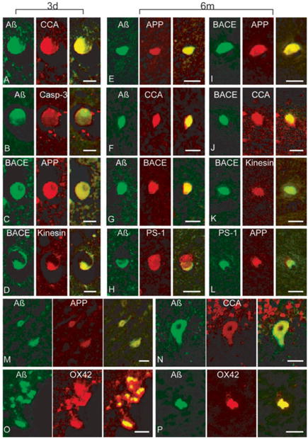
Fig. 12.

Co-localization of multiple proteins in cells and axons in swine post-TBI. Representative double-immunofluorescence photomicrographs demonstrating co-accumulations of proteins in damaged (a – l) axons, (m – n) neurons and (o – p) macrophages at 3 days and 6 months post-injury. Merged green and red fluorescence shown in yellow. In axon bulbs in the white matter, co-accumulation A (antibodies 6F3D and 13335/ Green) was found with CCA (249/Red) in (a) and (f), caspase-3 (P20/Red) in (b), BACE (BACE-2/Red) in (g), APP (22C11/Red) in (e), and PS-1 (PS-1/Red) in (h). Co-accumulation of BACE (Green) was found with APP (Red) in (c) and (i), kinesin (L1/Red) in (d) and (k), and CCA (Red) in (j). Co-accumulation of APP (Red) was found with PS-1 (Green) in (l) n neurons A (Green) co-accumulated with APP (Red) in (m) and CCA (Red) in (n). Macrophages demonstrated co-immunoreactivity of A (13335/Green) with OX42 (CD11b/Red) in (o) and (p). Scale bar = 25 μm. Reprinted with permission from ref. [92]
