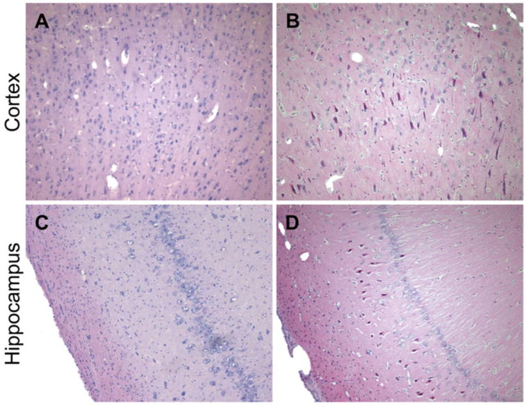
Fig. 13.

Neuronal degeneration following moderate-to-severe TBI in swine. H&E staining of the cerebral cortex and hippocampus in (a, c) sham pigs and (b, d) pigs subjected to closed-head rotational acceleration using the HYGE device. Neuronal degeneration, as shown by neuronal pyknosis, was observed at 7 days following sagittal plan rotation in the (b) cortex and (d) hippocampus
