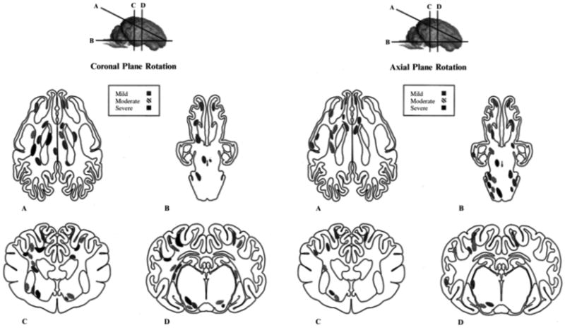
Fig 9.

Distribution of axonal pathology in pig model. Schematic representation of the distribution and severity of axonal injury following head coronal plane (left) and axial plane (right) rotation. Lines through the brain shown at the top of the figure demarcate anatomical regions of interest: frontal lobe, basal ganglia, and occipital lobe (a) brainstem through brain base (b) rostral thalamic level (c), and dorsal hippocampal level (d). Regions of axonal injury are shaded according to severity (mild, moderate, or severe). Reprinted with permission from ref. [98]
