
Figure 5.
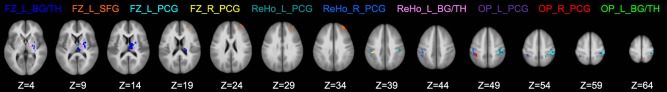
Brain regions that survived the SVM‐RFE analysis. Labels of different regions are in the same color as the region being labeled. FC: functional connectivity; ReHo: regional homogeneity; OP: overlapping regions between FC and ReHo; L: left; R: right; BG/TH: basal ganglia and thalamus; SFG: superior frontal gyrus; PCG: postcentral gyrus. [Color figure can be viewed at http://wileyonlinelibrary.com]