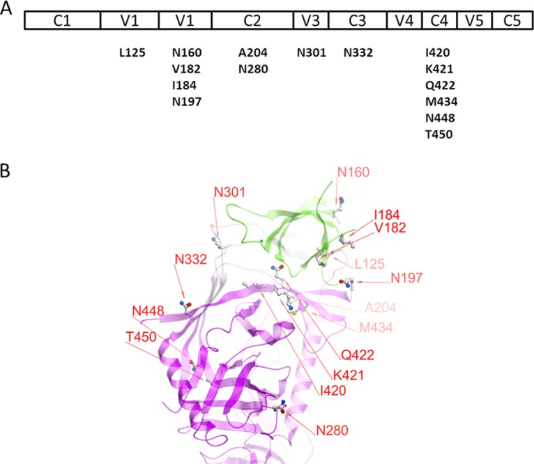
FIG 1.

Mutated residues of the JR-FL-JB pseudoviruses used in neutralization assays. (A) The various regions of gp120 are shown above the residues and positions in the wild-type virus that were mutated for this study. (B) Diagram of the positions of the mutations made in gp120. The V1V2 domain is shown in green, the gp120 core in magenta, and the V3 loop in gray ribbon representation. The image is based on PDB entry 4TVP, the crystal structure of the BG505 SOSIP trimer complexed with MAbs PGT122 and 35O22 (27). The numbering is based on the HxB2 reference sequence.
