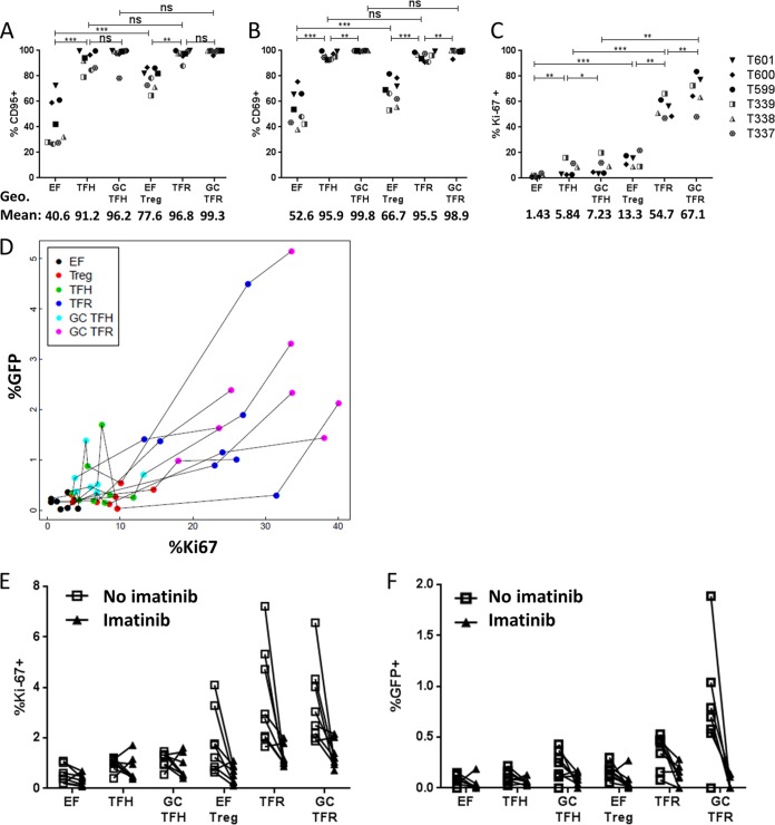
FIG 7.
Ki67 expression predicts GFP expression. (A to C) Disaggregated tonsil cells were stained for activation, memory, and proliferation markers at day 0 and immediately analyzed by flow cytometry, as indicated. Statistical significance was determined using repeated-measures one-way ANOVA. *, P < 0.05; **, P < 0.01; ***, P < 0.001; ns, not significant. Only pairwise comparisons of interest are shown. The overall P value was <0.05 (by ANOVA) for all. (D) A mixed-effects model was used to determine that the percentage of Ki67+ cells predicts the percentage of GFP+ cells for all T cell subsets investigated (n = 6; P < 0.0001). (E and F) Disaggregated tonsil cells were rested for 2 days with or without imatinib. At day 2, cells were split such that half were analyzed for Ki67 expression, and half were spinoculated with R5-tropic GFP reporter virus and cultured for another 2 days with or without imatinib (n = 6). The percentages of Ki-67+ and GFP+ cells were determined.

