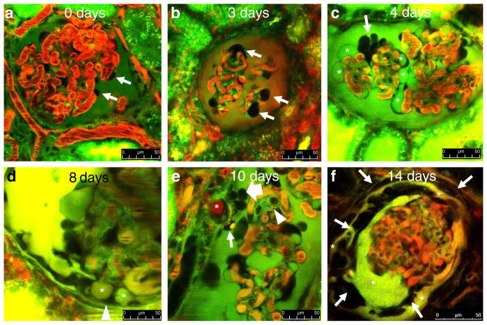
Fig. 2.
MPM imaging of the progression of glomerular pathology in vivo in a puromycin aminonucleoside (PAN)-induced model of FSGS in Munich-Wistar-Fromter rats. Red: intravascular space (plasma) marker Alexa 594-albumin given in iv bolus. Dark objects in red capillary plasma are red blood cells. Green/yellow: Lucifer yellow (LY) infused continuously into the carotid artery. In b, the nuclear stain Hoechst 33342 was also given iv to help the identification of various cells (green). The Bowman’s space is labeled by the freely filtered LY identifying podocytes (arrows) and parietal cells (which do not take up the dye) based on negative labeling. a Normal glomerular morphology is visible at baseline. b Podocytes with enlarged, balled-up cell body (arrows) appear 2–3 days after PAN treatment. c In addition to enlarged podocytes (arrow), numerous large cysts are visible in dark, unlabeled podocytes (asterisks) 4 days after PAN treatment. d Podocyte pseudocysts (asterisks) are in contact with the parietal Bowman’s capsule (arrowhead) at 8 days after PAN. e A fully formed adhesion (synechia) between the visceral and parietal Bowman’s capsules appearing 10 days after PAN, formed by an albumin (red)-containing pseudocyst (asterisk) and surrounded by several tightly packed cells (block arrow) and a podocyte containing green/yellow/red-labeled vesicles (arrow). Glomerular basement membranes are also labeled green by LY (arrowhead). f Multilayered Bowman’s capsule consisting of numerous cells (arrows) and LY-filled fluid spaces (asterisks) surrounds a barely perfused glomerulus (note the lack of red blood cells within glomerular capillaries) 14 days after PAN. Scale bars = 50 μm

