Fig. 4.
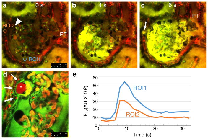
Time-lapse MPM imaging of misdirected filtration in vivo in PAN-treated Munich-Wistar-Fromter rat kidneys. Red: Plasma marker Alexa 594-albumin. Green/yellow: Freely filtered Lucifer yellow (LY) infused continuously into the carotid artery to label the primary filtrate. Images taken 12 days after PAN treatment. Glomerular leakage (visible red filtrate in Bowman’s space marked by ROI2 in a) and proximal tubule (PT) uptake of albumin are visible after PAN treatment. An adhesion (synechia) appears between the visceral and parietal Bowman’s capsule (arrowhead, a). Compared to the low background at time zero (a), bright LY fluorescence appears in glomerular capillaries and the Bowman’s space within 4 s of initiating carotid infusion (b). c At 6 s, high LY fluorescence appears at a specific focal region outside the glomerulus adjacent to the synechia (arrow, ROI1 in panel A), simultaneously with the initial filtration of LY into the Bowman’s space and PT lumen. d High albumin (red)-containing pseudocyst (asterisk) in a synechia appears to filter albumin into the periglomerular interstitium, which is taken up by numerous cells surrounding this focal region (arrows). e Line plot of LY fluorescence intensity changes (FLY) measured in two regions of interest (ROI1 in the Bowman’s space, and ROI2 outside the glomerulus) as indicated in a. Also, see Supplement movies 2–3 showing the same preparations. Scale bars = 50 μm

