Fig. 5.
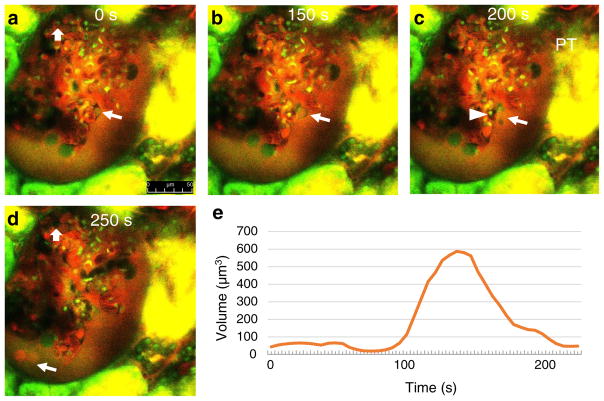
Time-lapse MPM imaging of podocyte shedding in vivo in PAN-treated Munich-Wistar-Fromter rat kidneys. Red: Plasma marker Alexa 594-albumin. Green/yellow: Lucifer yellow (LY) infused continuously into the carotid artery to label the primary filtrate. Hoechst 33342 identifies cell nuclei (green). Images taken 12 days after PAN treatment. Glomerular leakage of albumin is visible based on red filtrate in Bowman’s space around glomerular capillaries. At time zero (a), a small pseudocyst is present in the labeled podocyte (arrow), and an intact endothelial cell is visible in a glomerular capillary (block arrow). b Within 150 s, the same pseudocyst is significantly enlarged. c At 200 s, the pseudocyst is ruptured and shedding of cell debris is visible (arrow). A microthrombus (dark capillary mass, no red plasma) developed in the glomerular capillary directly adjacent to the injured podocyte (arrowhead). d Albumin-containing (red) vesicles appear shedding into the filtrate at 250 s. Note the absence of the same endothelial cell labeled in a, replaced by a microthrombus (block arrow). e Dynamic changes in the volume of the podocyte pseudocyst labeled in a–c. Also, see Supplement movie 4 showing the same preparation. Scale bar = 50 μm

