
Figure 1.
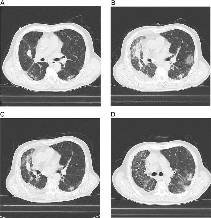
Chest computed tomography (CT) scans of the left upper lobe of the lung before and after initiating nivolumab. (A) No finding suggesting interstitial lung disease (ILD) before administration of nivolumab. (B) Appearance of the combination of ground‐grass nodule (GGN) and consolidation after eight cycles of nivolumab. (C) Improvement in CT findings after immunosuppressive treatment for 1 month; the GGN disappeared but the consolidation remained. (D) Re‐exacerbation of the combination of GGN and consolidation after tapering the dose of prednisolone to 30 mg daily.