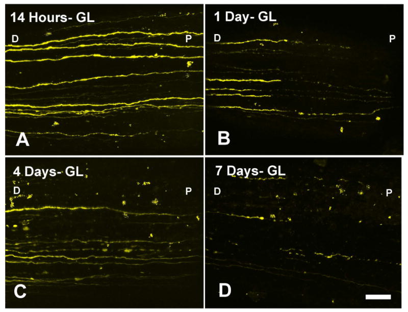
Figure 3.

RGC-YFP axons show progressive fragmenation, swelling, and loss after IOP elevation in the bead-induced model at (A) 14 hours, (B) 1 day, (C) 4 days, and (D) 1 week post chronic IOP elevation. Labels: D-distal to the eye, P-proximal to the eye (at the ONH) are used to indicate orientation (bar = 30 μm).
