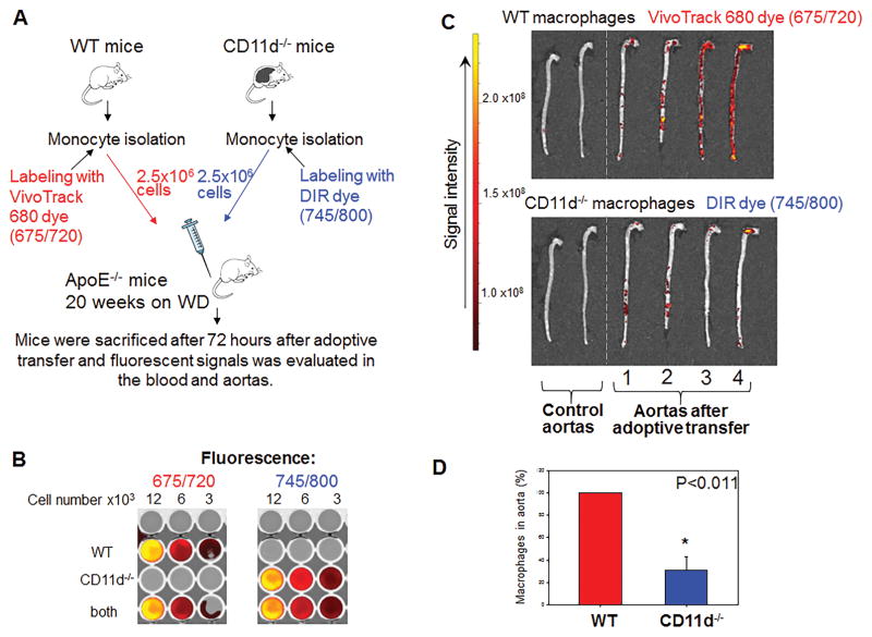
Fig. 6. Tracking the migration of adoptively transferred fluorescently-labeled monocytes to the atherosclerotic lesions of ApoE-deficient mice.
A. WT and CD11d-deficient monocytes were labeled with VivoTrack680 and DIR near infrared fluorescent dyes, correspondingly. An equal number of labeled WT and CD11d−/− cells was injected into the same ApoE−/− mouse. After 72 hours aortas from donor mice were isolated and fluorescence was measured using IVIC Spectrum CT Imaging system. B. The intensity of fluorescent signals and potential overlapping of dyes were normalized to the number of fluorescently labeled cells incubated in vitro by plating labeled monocytes in different concentrations in a 96-well plate. C. Two control aortas (no fluorescent cells injected) and four experimental aortas were evaluated using IVIC Spectrum CT Imaging system. D. The calculated result is based on the ratio between WT and CD11d−/− macrophages in atherosclerotic aortas. Data were plotted as the mean ± SEM. Statistical analysis was performed using Student’s t-test.

