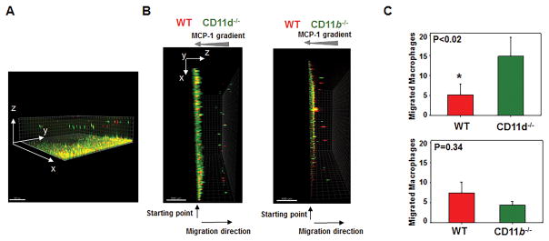
Fig. 7. CD11d-deficiency improves the migration of pro-inflammatory M1-activated macrophages in 3-D fibrin gels.
Migration of fluorescently labeled WT and CD11d-deficient (or CD11b-deficient) M1 macrophages in 3D fibrin toward an MCP-1 gradient (A, B). A. The example of 3-D view of migrated macrophages generated by collecting Z-stack images through fibrin matrix. B. Side view of generated 3-D images. WT and CD11d−/− peritoneal macrophages (B, left panel) or WT and CD11b−/− peritoneal macrophages (B, right panel) were activated in vitro for 3 days with IFNγ, labeled with PKH26 and PKH67 fluorescent dyes, respectively, and plated on 3D polymerized fibrin in transwell inserts. Migration of macrophages was stimulated by 30 nM MCP-1 added to the top of the gel. After 24 hours migrating cells were detected by a Leica Confocal microscope (Leica-TCS SP8) and the results were analyzed by IMARIS 8.0 software (C). Statistical analyses were performed using Student’s paired t-tests (n=4 per group). Scale bar – 500 μM.

