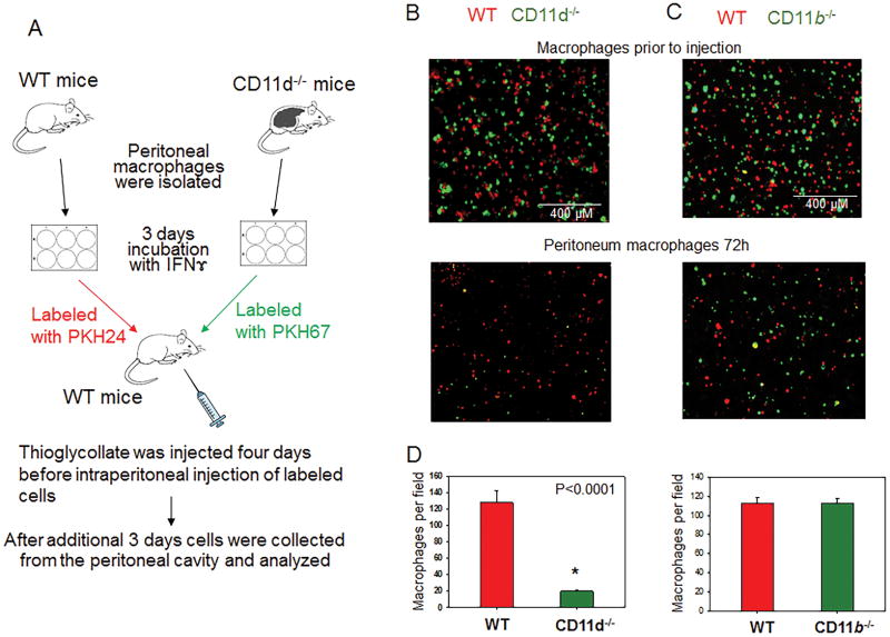
Fig. 8. CD11d-deficiency improves the efflux of pro-inflammatory M1-activated macrophages from the peritoneal cavity.
A. Peritoneal macrophages were isolated from WT and integrin-deficient mice at 3 days after injection of thioglycollate (TG) and labeled with PKH26 and PKH67 fluorescent dyes. Labeled WT and CD11d−/− or WT and CD11b−/− macrophages were mixed in a 1:1 ratio and further injected intraperitoneally into WT mice at 4 days after TG induced inflammation. The equal ratio of red and green macrophages before the injection was verified by sample cytospin preparation (B). 3 days later, peritoneal macrophages were harvested, cytospun and the percentages of red and green fluorescent macrophages were assessed by fluorescence microscopy using at least 9 fields of view per sample (n=6) (C). The quantification of the data was analyzed by using Image Analysis Software (EVOS, Thermo Fisher) (D). Statistical analysis was performed using Student’s paired t-tests.

