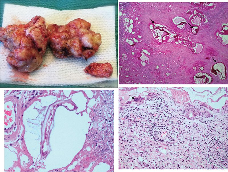
Fig. 5.

(Upper left) The gross pathologic specimen of the lesion removed during surgery. (Upper right) The section shows periodic acid-Schiff (PAS)-positive cuticle layer characteristic of Echinococcus multilocularis cysts (arrows). PAS stain. (Lower left) The cystic lesion with parasite surrounded by necrotic tissue. Hematoxylin and eosin stain. (Lower right) The surrounding brain tissue infiltrated by lymphocytes, plasmocytes, and multinucleated giant cells (arrow).
