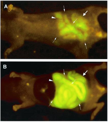
Figure 3.
Whole-body and intravital imaging of E. coli-GFP infection in the stomach, small intestine, and colon after gavage. (A) Whole-body image of E. coli-GFP infection in the stomach (arrowhead), the small intestine (fine arrows), and the colon (thick arrow) after multiple gavage of aliquots 3 × 10¹¹ E. coli-GFP. (B) Intravital image of E. coli-GFP infection in the stomach (arrowhead), the small intestine (fine arrows), and the colon (thick arrow) after multiple gavage of aliquots of 3 × 10¹¹ E. coli-GFP.

