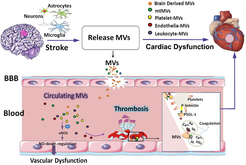
Figure 5. The role of microvesicles released by damaged astrocytes, neurons and microglia after stroke in mediating cardiac damage warrants future investigation.
Stroke induced elevation of circulating platelets and microvesicles (MVs) are associated with endothelial dysfunction and coagulation leading to thrombosis. MVs induce endothelial dysfunction by decreasing nitric oxide (NO) synthesis as the result of endothelial nitric oxide synthase (eNOS) function inhibition and an increase in caveolin-1. The outer leaflet of the MVs membrane contains two pro-coagulants: phosphatidylserine and tissue factor. MVs can bind to coagulation factors and promote their activation. In addition, MVs harbor tissue factor, which can initiate extrinsic coagulation pathways and promote the assembly of clotting enzymes leading to thrombin generation. At sites of vascular injury, P-selectin exposure by activated endothelial cells or platelets leads to the rapid recruitment of MVs bearing the P-selectin glycoprotein ligand-1 (PSGL-1). The interactions between PSGL-1 and platelet P-selectin are necessary to concentrate tissue factor activity at the thrombus.

