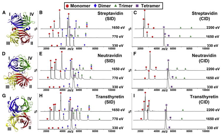
Figure 6.
Nano-electrospray SID (middle panel) and CID (right panel) mass spectra of the charge-reduced +11 precursor of (B and C) SA, (E and F) neutravidin, and (H and I) TTR at three separate collision energies. All fragments are labeled based on their corresponding peaks detected in IM. The precursor ion in each spectrum is indicated by a purple asterisk. Crystal structures of (A) SA (PDB: 1SWB), (D) neutravidin (PDB: 1VYO), and (G) TTR (PDB: 1F41) are shown in the left panel. Subunits I, II, III, and IV are also shown in blue, green, yellow, and red, respectively. Reproduced from Chemistry & Biology, Vol. 22, Quintyn, Q., Yan, J., Wysocki, V.H., Surface-Induced Dissociation of Homotetramers with D2 Symmetry Yields their Assembly Pathways and Characterizes the Effect of Ligand Binding, pp. 583–592, Copyright (2015), (ref. 142) with permission from Elsevier.

