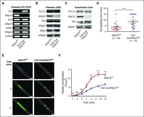
Figure 1.
Intravascular ERp72 is required for hemostasis and platelet accumulation into a growing thrombus. (A-C) Characterization of Tie2-Cre/ERp72fl/fl mice. (A) Platelet mRNA expression was evaluated by RT-PCR to demonstrate the absence of ERp72 mRNA. The mRNA expressions of other PDIs serve as control. Western blots of platelet (B) and endothelial (C) lysates using a polyclonal rabbit anti–ERp72 antibody and antibodies against PDI, ERp57, ERp46, and ERp5. Shown are the PLCγ2 loading controls for ERp72. Separate loading controls were run for ERp57, ERp5, and ERp72 with similar amounts of protein found in each sample (not depicted). Blots are representative of 3 separate experiments. (D) Tail bleeding times; ***P < .001, Student t test. (E and F) Incorporation of platelets into growing thrombus in ERp72fl/fl mice and Tie2-Cre/ERp72fl/fl mice was detected by Alexa 488 anti-CD41 using FeCl3-induced mesenteric arterial injury. Mean artery diameters were 125.2 ± 2.6 μm in ERp72fl/fl mice and 121.3 ± 2.1 μm in Tie2-Cre/ERp72fl/fl mice (P = not significant). (E) Images at 7, 9, and 12 minutes. Dotted lines mark the vessel wall. Scale bar, 200 μm. Images are original magnification ×100. (F) Composite of fluorescence intensity (FI) per area analyzed (FI/μm2) in ERp72fl/fl (n = 20 from 8 mice) and Tie2-Cre/ERp72fl/fl (n = 17 from 8 mice) mice; mean ± standard error of the mean (SEM), *P < .05, ***P < .001, Student t test.

