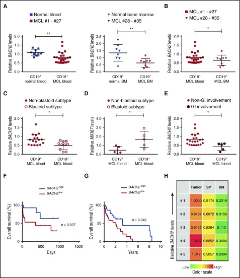
Figure 3.
Decreased BACH2 levels are associated with an unfavorable prognosis in MCL patients and promote tumor dispersal. (A) CD19+ B cells isolated from either apheresis blood samples (MCL patients, n = 27; healthy donors, n = 9) (left) or BM samples (MCL patients, n = 8; healthy donors, n = 8) (right) were used to measure the BACH2 mRNA levels using qRT-PCR. Each condition was run in triplicate with the values normalized to GAPDH. (B) BACH2 mRNA levels were downregulated in MCL cells isolated from BM (n = 8) compared with MCL isolated from blood samples (n = 27). (C) CD19+ cells isolated from MCL blastoid patients (n = 10) contained lower levels of BACH2 than CD19+ cells from MCL nonblastoid patients (n = 17). (D) MKI67 mRNA levels were measured in CD19+ B cells isolated from MCL blastoid subtypes (n = 5) or non-blastoid subtypes (n = 5). (E) CD19+ cells isolated from MCL patients with gastrointestinal (GI) dispersal (n = 5) contained lower levels of BACH2 compared with those from MCL patients without GI dispersal (n = 22). (F) The overall survival of MCL patients was significantly lower in BACH2low MCL patients than in BACH2high MCL patients (P = .027, Mantel-Cox curve analysis). BACH2high and BACH2low refer to the upper and lower 50% of BACH2 levels in MCL patients, respectively. (G) The overall survival of MCL patients was analyzed in Oncomine database using the upper and lower 25% of BACH2 levels in MCL patients with a cutoff of 10 years (P = .042, Mantel-Cox curve analysis). (H) Heatmap of BACH2 mRNA levels in subcutaneous tumor cells, SP, or BM from xenografts bearing MCL cells (n = 5, #1-#5). The fold change in expression compared with #1 xenograft tumor cells is indicated by the color intensity, with green representing reduced expression and red representing elevated expression. The results are shown as the mean ± SD. *P < .05; **P < .01 (Student t test).

