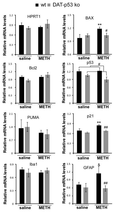
Fig 2.
Dopaminergic neuronal-specific p53 deletion results in differential gene transcriptional regulation in SN after MA binge exposure. Brain tissues were obtained 48 hours after MA binge challenge. qPCR analysis was conducted to determine the expression level of HPRT, p53, Bcl2, BAX, PUMA, p21, Iba1 and GFAP in DAT-p53KO and -WT mice. At this 48 hour time point, MA binge exposure induced upregulation of BAX, p21 and GFAP genes and these upregulations were attenuated in DA-p53KO mice. Data are shown as mean ± SEM. * and ** indicates p<0.05 or p<0.01, & indicates p=0.08 in MA binge exposed WT mice compared to saline injected mice. #, or ## indicates p<0.05 or p<0.01 in MA injected DAT-p53KO vs. -WT mice, n=8–10 for each group.

