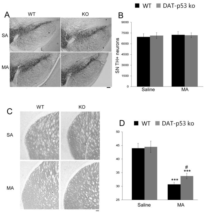
Figure 5.
Dopaminergic specific p53 deletion sustained the protection of DA terminals within the striatum, as evaluated at 10 days following MA binge exposure. (A) No loss of TH positive neurons was evident within the SN of MA injected WT or DA-p53KO mice, (B) which was confirmed by unbiased stereological counts. (C) MA binge led to a decrease in striatal TH immunoreactivity density in both WT and DA-p53KO mice when evaluated 10 days after MA binge. Notable, however, DA-p53KO mice continue to demonstrate a higher density of TH-immunoreactivity within the striatum vs. -WT mice at 10 days after MA binge exposure (D). *** indicates p<0.001 compared to saline injected mice and # indicates p<0.05 WT vs KO mice=7–8 for each group). (Calibration bar =100 um).

