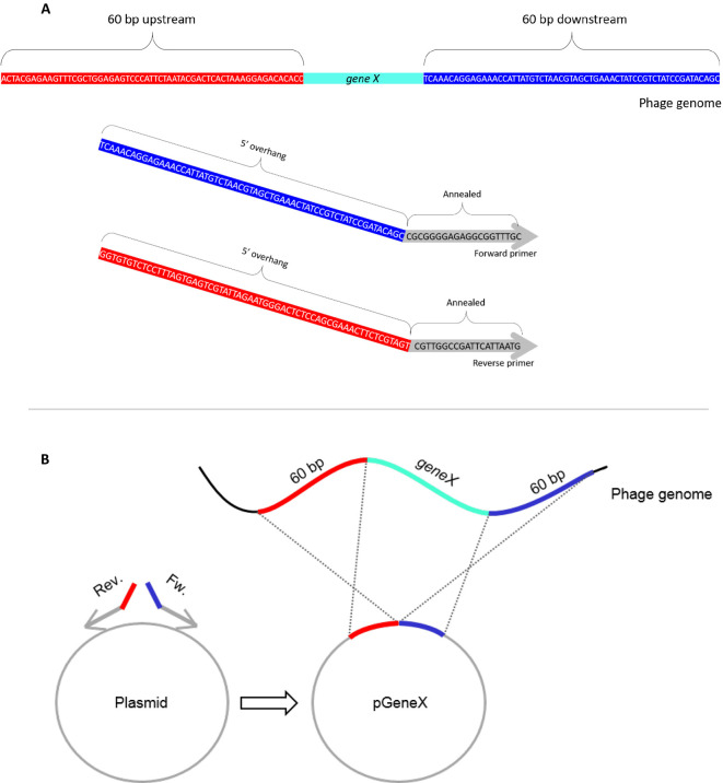
Figure 1. Schematics of the pGeneX plasmid.
A. An example of forward and reverse primers encoding sequences annealing to the pUC19 backbone (gray) with 60 bp 5’ overhang sequence identical to the upstream (red) and downstream (blue) flanking sequences of geneX. B. PCR amplification with these primers on the plasmid and self-ligation yield the pGeneX plasmid.

