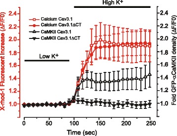
Fig. 5.

Cav3.1-mediated activation of αCaMKII depends on an intact Cav3.1 C-terminus. tsA-201 cells are transfected with Cav3.1 or a Cav3.1 mutant lacking the C-terminus (Cav3.1ΔCT) to remove a site for preassociation of CaM with the Cav3.1 channel and preloaded with 2 μM X-rhod-1 to detect changes in [Ca]i. Cells are maintained at rest in low [K]o (1 mM) or exposed to 10 min of high [K]o (50 mM), with all medium containing 30 μM Cd2+ to block HVA calcium channels. The extent and timecourse of a shift in GFP-αCaMKII distribution from diffuse to aggregates in the cytoplasm is compared to that of a change in fluorescent intensity of X-rhod-1 relative to the mean of control baseline (ΔF/F0). High [K]o increased [Ca]i to an equivalent relative level in cells expressing either Cav3.1 or Cav3.1ΔCT but GFP-αCaMKII aggregation is blocked in cells expressing the Cav3.1ΔCT mutant. The distribution of GFP-αCaMKII is measured as density for at least 25–34 ROIs from 4 to 5 plates. Values are mean ± SEM derived from n = 4–5 plates with 25–34 ROIs
