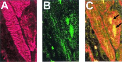
Figure 5.
Confocal microscopic colocalization of BrdUrd and of sarcomeric α-actinin within the nuclei of MRL vetricular cardiomyocytes 15 days after injury. (A) The immunocytochemical detection sarcomeric α-actinin in cardiomyocytes by using a Cy5-conjugated secondary antibody whose fluorescent infrared emission was visualized in a false-red color. (B) The immunocytochemical detection of BrdUrd, by using an FITC-conjugated secondary antibody; (C) the combined images of A and B can be seen. The arrows indicate the BrdUrd-labeled nuclei.

