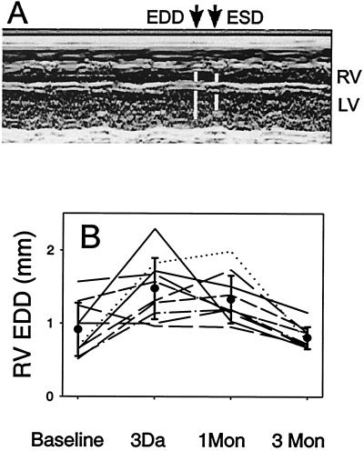
Figure 6.
Echocardiography of injured hearts. (A) An M-mode image from an MRL mouse 3 days after injury showing the ventricular chamber (RV, Upper; LV, Lower) dimensions throughout several cardiac cycles. The time points identified by the arrows allow the measurement of the end diastolic dimension (EDD, Left) and end systolic dimension (ESD, Right). The RV EDD is indicated by the upper left white bar. (B) The time course of right ventricular end diastolic diameter at base line, early after injury, 1 and 3 months after injury is presented. Individual lines show the response of individual MRL mice at each time point. The mean measurements are indicated (●), and the error bars are the standard deviation. The MRL mouse right ventricles dilate early in response to injury and recover by shrinking to their original size by 3 months.

