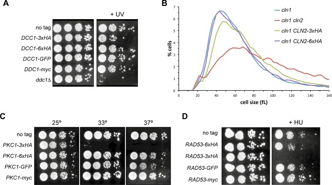
Fig 4. Functional analysis of proteins labelled with different tags.
(A) 10-fold serial dilutions from exponentially growing cultures of wild-type (W303-1a), DDC1-3xHA (JCY1701), DDC1-6xHA (JCY1825), DDC1-GFP (JCY1661) and DDC1-myc (JCY1887) strains were spotted onto YPD medium and exposed to UV radiation (40 J/m2). Plates were incubated at 25°C for 4 days. (B) Cell size distribution in exponentially growing cultures of cln1 (JCY275), cln1 cln2 (JCY847), cln1 CLN2-3xHA (JCY929) and cln1 CLN2-6XHA (JCY1960) in complete SD medium. (C) 10-fold serial dilutions from exponentially growing cultures of wild-type (W303-1a), PKC1-3xHA (JCY2033), PKC1-6XHA (1891), PKC1-GFP (JCY1511) and PKC1-myc (JCY1916) were spotted onto YPD medium. Plates were incubated at 25°C, 33°C or 37°C for 3 days. (D) 10-fold serial dilutions from exponentially growing cultures of wild-type (W303-1a), RAD53-6xHA (JCY1901), RAD53-3XHA (JCY1905), RAD53-GFP (JCY1903) and RAD53-myc (JCY1907) were spotted onto YPD medium containing 200 mM hydroxyurea. Plates were incubated at 25°C for 3 days.

