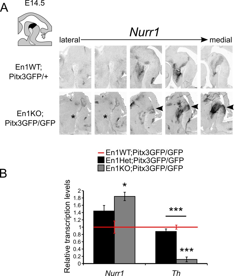
Fig 4. Nurr1-positive mdDA neurons are present in double En1KO;Pitx3GFP/GFP animals at E14.5.
(A) Schematic, sagittal section of the embryonic mouse brain, mdDA area is indicated in black. Lateral to medial sections of in situ hybridization experiments for Nurr1 at E14.5 in En1WT;Pitx3GFP/+ and En1KO;Pitx3GFP/GFP midbrains. Asterisk indicates diminished expression of Nurr1 in lateral sections, whilst in (para)medial sections Nurr1 is still present in the midbrain and is ectopically extended caudally (arrowheads). (B) Quantitative PCR on FAC-sorted mdDA neurons demonstrates elevated level of Nurr1 in the En1KO;Pitx3GFP/GFP midbrain, compared to En1WT;Pitx3GFP/GFP (* = P<0.05, n = 3/4). The expression of Th expression is significantly down-regulated in the En1KO;Pitx3GFP/GFP midbrain, compared to En1WT;Pitx3GFP/GFP (*** = P<0.01, n = 3/4).

