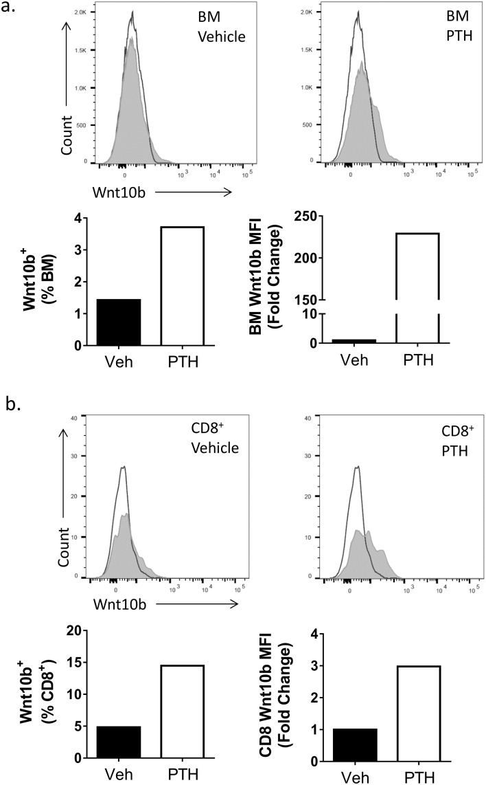
Fig 1. Detection of Wnt10b by flow cytometry.
Bone marrow (BM) was isolated from the femur of a male C57BL/6 mouse (n = 1) and cultured ± 50nM PTH for 3 hours. Cells were stained for Wnt10b and analyzed by flow cytometry, gating on bone marrow and CD8+ T cells. In the bone marrow population (a) a shift in peak was observed for the Wnt10b stained cells over the isotype control corresponding to approx. 1.5% of vehicle treated cells. PTH increased the number of Wnt10b+ BM cells to 3.5% and the MFI greater than 200-fold. Gating on the CD8+ T cells (b) in the vehicle treated group identified approximately 5% were Wnt10b+. Following PTH treatment this increased 3-fold.

