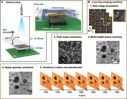
Fig. 1. Lens-free on-chip microscopy setup and image processing steps.

(A) Schematic of the lens-free on-chip imaging setup. The cleared tissue is loaded in a polydimethylsiloxane (PDMS)/glass chamber filled with a refractive index matching solution. A sealant is applied on the sides to avoid evaporation and leakage. RIMS, refractive index matching solution; CMOS, complementary metal-oxide semiconductor. (B) Lens-free image processing workflow is outlined.
