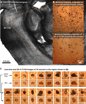
Fig. 5. Lens-free 3D imaging of a cleared, DAB-stained, 200-μm-thick mouse brain tissue.

(A) Full FOV lens-free hologram. (B) A zoomed-in region corresponding to a 20× microscope objective FOV. MIP images of the lens-free pseudocolored z-stack and the scanning microscope’s z-stack [obtained with a 20× objective (NA = 0.75)] are presented. (C) Comparison of lens-free images of 19 neurons against the images obtained with a 20× objective lens (NA = 0.75).
