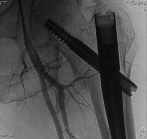
Figure 5.

Conventional arteriography: The bone spike of the lesser trochanter appears in contact with deep femoral artery. In our case, a reasonable explanation for the formation of the vascular lesion could be the closeness between lesser trochanter and deep femoral artery
