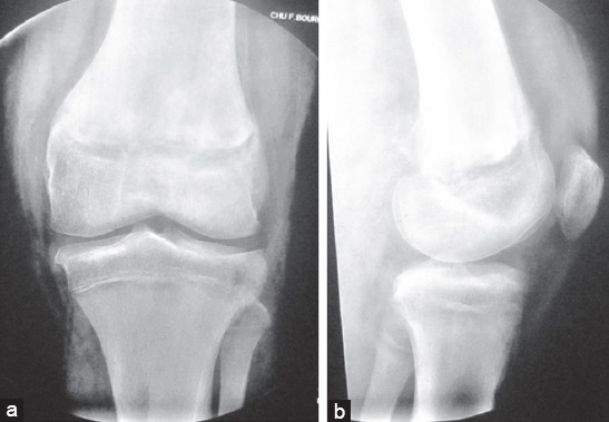
Figure 1.

(a and b) Pre-operative plain radiograph of the left knee showing a Salter and Harris type I physeal fracture of the distal femur with an important posterior displacement of the epiphysis together with a displaced type III physeal fracture of the proximal tibia interesting the lateral tibial plateau (1a front view, 1b lateral view)
