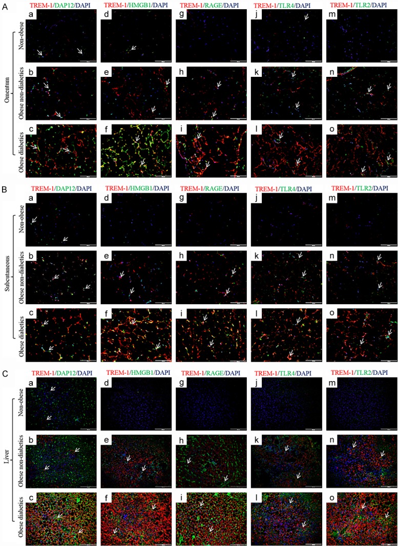
Figure 3.

Immunofluorescence staining for TREM-1, DAP12, HMGB1, RAGE, TLR-4 and TLR-2 in the biopsy samples of study subjects. Dual fluorescence staining was done using anti-human TREM-1 (Alexa 594-Red) and DAP12, HMGB1, RAGE, TLR-4 and TLR-2 (Alexa 488-green) antibodies to co-localize TREM-1 with DAP12, HMGB1, RAGE, TLR-4 and TLR-2 respectively, counterstained with DAPI. Negative controls were run by using isotypes for each fluorochrome. A: Immunofluorescence staining in omentum biopsy samples. B: Immunofluorescence staining in subcutaneous biopsy samples. C: Immunofluorescence staining in liver biopsy samples. (N=5 non-obese; 24 obese non-diabetics; 22 obese diabetics).
