
Figure 5.
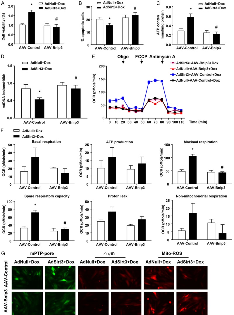
AAV-Bnip3 completely abolished the cardioprotective effect of Sirt3 on Dox-treated cardiomyocytes in vitro. (A) Quantification of cell viability by CCK8 assay. (B) Flow cytometry analysis was used to detect the level of apoptosis of cardiomyocytes via Ann-V (x axis) and PI (y axis) staining. (C) ATP content of cardiomyocytes under the same conditions (mg/g protein). (D) Quantitative PCR analysis was used to assess mtDNA damage. (E) OCR was measured with a Seahorse metabolic analyzer. Oligomycin (1 µM), FCCP (1 µM), and rotenone (1 µM) combined with antimycin (1 µM) were added sequentially to AdNull + AAV-Control/Bnip3- and AdSirt3 + AAV-Control/Bnip3-infected cardiomyocytes after Dox treatment. (F) Histograms showing basal respiration, ATP production, maximal respiration, spare respiratory capacity, proton leak, and non-mitochondrial respiration data for (E). (G) Fluorescence microscopy of AdNull + AAV-Control/Bnip3- and AdSirt3 + AAV-Control/Bnip3-infected cardiomyocytes after Dox treatment for mPTP opening (left), mitochondrial ΔΨm (center), and ROS (right). Data are presented as mean ± SEM. *P<0.05, **P<0.01, **P<0.001 versus the corresponding vehicle AdNull group; #P<0.05 versus the corresponding AAV-Control group.